
Routine eye exams are important, regardless of your age or physical health. During a complete eye exam, your eye doctor will not only determine your prescription for eyeglasses or contact lenses, but will also check your eyes for common eye diseases, assess how your eyes work together as a team and evaluate your eyes as an indicator of your overall health.
Eye Exams For the Whole Family
Need an Eye Exam to Update Your Prescription?
A comprehensive eye exam includes a number of tests and procedures to examine and evaluate the health of your eyes and the quality of your vision. These tests range from simple ones, like having you read an eye chart, to complex tests, such as using a high-powered lens to examine the health of the tissues inside of your eyes.
Eye care experts recommend you have a complete eye exam every year to assess your risk for potentially damaging eye conditions such as cataracts, glaucoma, and macular degeneration, as well as to keep on top of any changes in vision you may be experiencing.
Your Comprehensive Eye Exam
Your eyes are one of the most complex organs in your body. A comprehensive eye exam to assess your visual system and eye health involves a number of different of tests. Unlike a simple vision screening, which only assesses your vision, a comprehensive eye exam includes a battery of tests in order to do a complete evaluation of the health of your eyes and your vision.
The tests that you will undergo in a comprehensive eye examination may vary from eye doctor to eye doctor but here are are some common exams that you may encounter:
Patient Background and History
One of the most important parts in a comprehensive eye exam is your patient health history. This information will alert your doctor to any conditions that should be monitored closely, such as an allergy to any medications, current or family history of systemic or eye pathology or environmental conditions that could be affecting your vision or eye health. This will also help your doctor to determine any preventative eye care measures that are relevant to keep your eyes healthy for years to come.
Visual Acuity
Visual acuity is a measurement of your vision using an eye chart, the Snellen Eye Chart. In this test the patient is seated at a standard distance and is asked to read letters or symbols of various sizes, which get smaller as you move down the chart. The results are the familiar ratio of 20/20, 20/40 etc. which is a comparison of your vision compared to the average person with good vision, which is typically 20/20. For example, a patient that has 20/40 vision, can only see at 20 feet what the normal person can see from a distance of 40 feet. This test is a preliminary test of how clearly you are seeing in each eye but it does not give you a prescription for corrective lenses.
Refraction
Those who don’t have 20/20 vision have what is referred to in most cases as a “Refractive Error.” The patient may have nearsightedness, farsightedness, astigmatism or other eye conditions that prevent the patient from seeing 20/20. A refraction will tell the doctor which prescription lenses will correct your eyesight to achieve 20/20 vision or whichever amount your vision is correctable to.
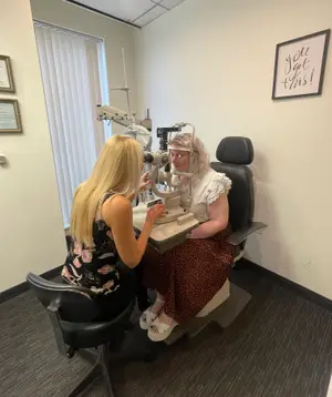
We are committed to providing you with the most precise vision correction possible. During your comprehensive eye exam, your doctor will determine your prescription using a phoropter. For maximum accuracy and patient comfort, all three of our exam lanes are equipped with state-of-the-art, fully automated digital auto-phoropters. This technology allows us to seamlessly evaluate your vision and quickly pinpoint your ideal prescription.
Retinoscopy
Retinoscopy is a test that allows the doctor to obtain an approximate prescription for eyeglasses. In this test the doctor uses a hand-held instrument called a retinoscope that shines a light into the patient’s eye. The doctor then analyzes the reflex of the light from the patient’s eye to determine the patient’s prescription for glasses.
An instrument called a phoropter is something most patients associate with an eye exam. This space age appearing instrument, positioned in front of the patient’s face during the eye exam, gives the doctor the ability to determine the patient’s focusing ability as well as their eye alignment. The phoropter also determines which, out of the hundreds and hundreds of potential eyeglass prescriptions, will help the patient see as clear as possible. Using the phoropter, the doctor will ask the patient which series of lenses makes their vision the clearest.
While retinoscopy is quite effective for children and nonverbal patients, there are now a number of computerized or automated instruments available today to help doctors accurately determine a patient’s eyeglass prescription.
Eye Focusing and Eye Teaming Tests
During the comprehensive eye exam, your eye doctor will also want to test how your eyes function individually and together from a mechanical perspective. In order to see clearly and comfortably, your eyes need to work together as a team.
Eye Health
The final and most important aspect of a comprehensive eye exam is a check of your overall eye health. These tests (below) are done to identify any eye conditions or diseases, both inside the eye as well as the external parts of the eye, that could affect your vision and general health.
Slit Lamp Test
The slit lamp or biomicroscope is an instrument that allows the doctor to examine the internal and external parts of the eye in detail, such as the conjunctiva, iris, lens, cornea, retina and the optic nerve. The patient rests their forehead and chin on a headrest to stabilize the head, while the doctor looks into the eye with the slit lamp microscope, which is magnified with a high-intensity light. A slit lamp test enables the doctor to evaluate the eyes for signs of normal aging and eye pathology, such as conjunctivitis, cataracts, macular degeneration or retinal detachment. Early diagnosis and treatment of eye diseases are essential for preventing vision loss.
Tonometry
Tonometry is a test to detect glaucoma by measuring the pressure inside your eye or IOP (intraocular pressure). Glaucoma can cause vision loss and even blindness if the IOP in the eye is too high and damages the optic nerve.
The applanation tonometer, typically attached to a slit lamp, is one of the most common instruments used to measure the pressure in the eye. Prior to doing this test the doctor will numb the patient’s eyes using an anesthetic, before gently applanating (putting pressure on) the patient’s cornea to measure the pressure in the eye.
Optomap
Dr. Norton considers the Optomap Retinal Exam as an essential part of your eye health exam. The Optomap Retinal Exam captures a digital image that is a unique as your fingerprint. It provides the doctor with an ultra wide field view to examine the health the most important structures of your eye, including your retina, macula, and optic nerve. Many eye problems can develop without you realizing it. You may not even notice any change in your sight. Diseases such as macular degeneration, glaucoma, retinal tears or detachments, and other health problems such as diabetes and high blood pressure can be seen with a thorough exam of the retina. An Optomap Retinal Exam with the newest technology using the Optos Monaco Pro provides: A digital image to show a healthy eye or detect disease
- Comfortable and quick image capture
- Non-invasive examination without any side effects
- A full view of the retina, giving your doctor a more detailed view that she can get by other methods
- The opportunity for you to view and discuss the Optomap image of your eye
- A permanent record for future comparison
The Optomap technology does not require pupil dilation. You can resume normal activities immediately since there is no blurred vision or light sensitivity as occurs with dilation. However, in some cases, dilation may also be necessary in addition to the Optomap to provide a thorough examination. The decision to dilate or not is a medical decision made by the doctor based on your history and examination findings. The Optomap Retinal Exam is fast, easy, and comfortable for all ages. To have the exam, you simply look into the device one eye at a time and you will see a quick flash of light similar to a camera flash. The Optomap image is then available immediately for the doctor to review and evaluate. In some cases, the doctor will recommend a dilated fundus exam in addition to retinal imaging. This is decided on an individual basis depending on ocular, medical, and family history as well if you there are any concerning symptoms or exam findings.